Oregon State Flag An official website of the State of Oregon »

Oregon Health Authority

Parasite Images from the Oregon State Public Health Laboratory


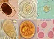
These microphotographic images of parasites were taken at the Oregon State Public Health Laboratory. The OSPHL Parasitology Department is equipped with a QImaging MicroPublisher 3.3 RTV high resolution Digital Camera mounted on a Leica DMLS microscope.

This equipment is used in conjunction with the Division of Parasitic Diseases (DPDx) at the Centers for Disease Control & Prevention (CDC) to aid in the rapid diagnosis of unusual or difficult parasitic diseases using digital images and the Internet.

Click on a thumbnail image below to see a larger version and brief description of that particular parasite.

Balantidium image


This parasite is a ciliate protozoan. The trophozoites measure 50 to 200 microns. Viable organisms in unfixed stool samples are very active moving across the field of view in a rotary motion.

This microphotographic image was taken at the Oregon State Public Health Laboratory. The OSPHL Parasitology Department is equipped with a Leica DC 200 digital camera mounted on a Leica DMLS microscope. This equipment is used in conjunction with the Division of Parasitic Diseases (DPDx) at the Centers for Disease Control & Prevention (CDC) to aid in the rapid diagnosis of unusual or difficult parasitic diseases using digital images and the Internet. ​


Baylisascaris Egg image


Baylisascaris Egg: This egg came from a racoon, but the same results can sometimes be seen in Ascaris eggs from human samples .

This microphotographic image was taken at the Oregon State Public Health Laboratory. The OSPHL Parasitology Department is equipped with a Leica DC 200 digital camera mounted on a Leica DMLS microscope. This equipment is used in conjunction with the Division of Parasitic Diseases (DPDx) at the Centers for Disease Control & Prevention (CDC) to aid in the rapid diagnosis of unusual or difficult parasitic diseases using digital images and ​the Internet.

Blastocystis hominis image


Blastocystis hominis: This organism measures 4 to 35 microns. The possible pathogenicity of this organism is a matter of controversy.

This microphotographic image was taken at the Oregon State Public Health Laboratory. The OSPHL Parasitology Department is equipped with a Leica DC 200 digital camera mounted on a Leica DMLS microscope. This equipment is used in conjunction with the Division of Parasitic Diseases (DPDx) at the Centers for Disease Control & Prevention (CDC) to aid in the rapid diagnosis of unusual or difficult parasitic diseases using digital images and the Internet.

Chilomastix mesnili image

​Chilomastix mesnili: is a nonpathogenic flagellate protozoan. The lemon shaped uninucleate cysts can measure from 6 to 10 microns.

This microphotographic image was taken at the Oregon State Public Health Laboratory. The OSPHL Parasitology Department is equipped with a Leica DC 200 digital camera mounted on a Leica DMLS microscope. This equipment is used in conjunction with the Division of Parasitic Diseases (DPDx) at the Centers for Disease Control & Prevention (CDC) to aid in the rapid diagnosis of unusual or difficult parasitic diseases using digital images and the Internet.

​​

Cryptosporidium image

Cryptosporidium oocysts: These spherical oocysts measure 4 to 6 microns and stain acid fast positive with modified acid fast stains.

This microphotographic image was taken at the Oregon State Public Health Laboratory. The OSPHL Parasitology Department is equipped with a Leica DC 200 digital camera mounted on a Leica DMLS microscope. This equipment is used in conjunction with the Division of Parasitic Diseases (DPDx) at the Centers for Disease Control & Prevention (CDC) to aid in the rapid diagnosis of unusual or difficult parasitic diseases using digital images and the Internet.

Entamoeba coli image


Entamoeba coli
: These cysts measure 15 to 50 microns in diameter. When mature they usually contain 8 nuclei. This organism is nonpathogenic.

This microphotographic image was taken at the Oregon State Public Health Laboratory. The OSPHL Parasitology Department is equipped with a Leica DC 200 digital camera mounted on a Leica DMLS microscope. This equipment is used in conjunction with the Division of Parasitic Diseases (DPDx) at the Centers for Disease Control & Prevention (CDC) to aid in the rapid diagnosis of unusual or difficult parasitic diseases using digital images and the Internet.

Entamoeba hartmanni Image

Entamoeba hartmanni: These cysts can measure from 5 to 10 microns. They can contain up to four nuclei. This organism is nonpathogenic.

This microphotographic image was taken at the Oregon State Public Health Laboratory. The OSPHL Parasitology Department is equipped with a Leica DC 200 digital camera mounted on a Leica DMLS microscope. This equipment is used in conjunction with the Division of Parasitic Diseases (DPDx) at the Centers for Disease Control & Prevention (CDC) to aid in the rapid diagnosis of unusual or difficult parasitic diseases using digital images and the Internet.

Entamoeba histolytica/dispar image

Entamoeba histolytica/dispar: Cysts measure 10 to 20 microns. The four nuclei in this cyst is typical of these species. To count all four nuclei requires focusing up and down through the cyst. This changing of focal planes also helps show the chromatoid body as a cigar shaped object with rounded ends. 

This microphotographic image was taken at the Oregon State Public Health Laboratory. The OSPHL Parasitology Department is equipped with a Leica DC 200 digital camera mounted on a Leica DMLS microscope. This equipment is used in conjunction with the Division of Parasitic Diseases (DPDx) at the Centers for Disease Control & Prevention (CDC) to aid in the rapid diagnosis of unusual or difficult parasitic diseases using digital images and the Internet.

Wet mount cyst Giardia lamblia image

Giardia lamblia
 (wet mount cyst):  This organism is a pathogenic flagellate. This animated GIF shows the view one would see as they focus up and down through the organism’s cyst stage in a wet mount preparation.

This microphotographic image was taken at the Oregon State Public Health Laboratory. The OSPHL Parasitology Department is equipped with a Leica DC 200 digital camera mounted on a Leica DMLS microscope. This equipment is used in conjunction with the Division of Parasitic Diseases (DPDx) at the Centers for Disease Control & Prevention (CDC) to aid in the rapid diagnosis of unusual or difficult parasitic diseases using digital images and the Internet. 

Trichrome cyst Giardia lamblia image


Giardia Lamblia - Trichrome cyst
This is a trichrome stain of the cyst stage of Giardia lamblia. The cysts measure 8 to 19 microns.

This microphotographic image was taken at the Oregon State Public Health Laboratory. The OSPHL Parasitology Department is equipped with a Leica DC 200 digital camera mounted on a Leica DMLS microscope. This equipment is used in conjunction with the Division of Parasitic Diseases (DPDx) at the Centers for Disease Control & Prevention (CDC) to aid in the rapid diagnosis of unusual or difficult parasitic diseases using digital images and the Internet.

Wet mount trophozoite - Giardia lamblia

Wet mount trophozoite - Giardia lambliaThis organism is a pathogenic flagellate. This animated GIF shows the view one would see as they focus up and down through the organism’s trophozoite stage in a wet mount preparation.

This microphotographic image was taken at the Oregon State Public Health Laboratory. The OSPHL Parasitology Department is equipped with a Leica DC 200 digital camera mounted on a Leica DMLS microscope. This equipment is used in conjunction with the Division of Parasitic Diseases (DPDx) at the Centers for Disease Control & Prevention (CDC) to aid in the rapid diagnosis of unusual or difficult parasitic diseases using digital images and the Internet.

West mount cyst: Giardia lamblia

Wet mount cyst Giardia lamblia: The mature cysts of this organism contain four nuclei. Intracytoplasmic fibrils are visible in this iodine stained wet mount.

This microphotographic image was taken at the Oregon State Public Health Laboratory. The OSPHL Parasitology Department is equipped with a Leica DC 200 digital camera mounted on a Leica DMLS microscope. This equipment is used in conjunction with the Division of Parasitic Diseases (DPDx) at the Centers for Disease Control & Prevention (CDC) to aid in the rapid diagnosis of unusual or difficult parasitic diseases using digital images and the Internet.

Hookworm egg

​Hookworm egg
These eggs have the characteristic thin clear shell.

This microphotographic image was taken at the Oregon State Public Health Laboratory. The OSPHL Parasitology Department is equipped with a Leica DC 200 digital camera mounted on a Leica DMLS microscope. This equipment is used in conjunction with the Division of Parasitic Diseases (DPDx) at the Centers for Disease Control & Prevention (CDC) to aid in the rapid diagnosis of unusual or difficult parasitic diseases using digital images and the Internet.


Hymenolepis-nana (Dwarf tapeworm) image


Hymenolepis-nana (Dwarf tapeworm): These eggs measure 30 to 47 microns and contain a six hooked oncosphere..

This microphotographic image was taken at the Oregon State Public Health Laboratory. The OSPHL Parasitology Department is equipped with a Leica DC 200 digital camera mounted on a Leica DMLS microscope. This equipment is used in conjunction with the Division of Parasitic Diseases (DPDx) at the Centers for Disease Control & Prevention (CDC) to aid in the rapid diagnosis of unusual or difficult parasitic diseases using digital images and the Internet.

Iodamoeba beutschlii image

Iodamoeba beutschlii: The cysts of this nonpathogenic amoeba measure from 5 to 20 microns.

This microphotographic image was taken at the Oregon State Public Health Laboratory. The OSPHL Parasitology Department is equipped with a Leica DC 200 digital camera mounted on a Leica DMLS microscope. This equipment is used in conjunction with the Division of Parasitic Diseases (DPDx) at the Centers for Disease Control & Prevention (CDC) to aid in the rapid diagnosis of unusual or difficult parasitic diseases using digital images and the Internet.

Morel spore

Morel spore: This non-parasitic artifact was seen in moderate numbers in a patient that had recently eaten morel mushrooms.

This microphotographic image was taken at the Oregon State Public Health Laboratory. The OSPHL Parasitology Department is equipped with a Leica DC 200 digital camera mounted on a Leica DMLS microscope. This equipment is used in conjunction with the Division of Parasitic Diseases (DPDx) at the Centers for Disease Control & Prevention (CDC) to aid in the rapid diagnosis of unusual or difficult parasitic diseases using digital images and the Internet.

Nanophyetus species 

Nanophyetus species: This parasite is rarely seen. It can be acquired from eating undercooked fish. Nanophyetus species have been found in North America and from a region in Siberia.

This microphotographic image was taken at the Oregon State Public Health Laboratory. The OSPHL Parasitology Department is equipped with a Leica DC 200 digital camera mounted on a Leica DMLS microscope. This equipment is used in conjunction with the Division of Parasitic Diseases (DPDx) at the Centers for Disease Control & Prevention (CDC) to aid in the rapid diagnosis of unusual or difficult parasitic diseases using digital images and the Internet.​

Plasmodium vivax 

Plasmodium vivax:

This image shows two of the stages seen with this species of malaria.

This microphotographic image was taken at the Oregon State Public Health Laboratory. The OSPHL Parasitology Department is equipped with a Leica DC 200 digital camera mounted on a Leica DMLS microscope. This equipment is used in conjunction with the Division of Parasitic Diseases (DPDx) at the Centers for Disease Control & Prevention (CDC) to aid in the rapid diagnosis of unusual or difficult parasitic diseases using digital images and the Internet.​

Roundworm egg comparison 

Roundworm egg comparison: This image provides a side by side comparison of the eggs of three different roundworm species.

This microphotographic image was taken at the Oregon State Public Health Laboratory. The OSPHL Parasitology Department is equipped with a Leica DC 200 digital camera mounted on a Leica DMLS microscope. This equipment is used in conjunction with the Division of Parasitic Diseases (DPDx) at the Centers for Disease Control & Prevention (CDC) to aid in the rapid diagnosis of unusual or difficult parasitic diseases using digital images and the Internet.​​

Strongyloides stercoralis 

Strongyloides stercoralis: This is the rhabditiform larvae of this parasite. This stage is seen in the stools of infected patients. The egg stage is rarely seen.

This microphotographic image was taken at the Oregon State Public Health Laboratory. The OSPHL Parasitology Department is equipped with a Leica DC 200 digital camera mounted on a Leica DMLS microscope. This equipment is used in conjunction with the Division of Parasitic Diseases (DPDx) at the Centers for Disease Control & Prevention (CDC) to aid in the rapid diagnosis of unusual or difficult parasitic diseases using digital images and the Internet.​

Trichuris trichiura (Whipworm) 

Trichuris trichiura (Whipworm): The eggs of this parasite measure 50 to 55 microns in length.

This microphotographic image was taken at the Oregon State Public Health Laboratory. The OSPHL Parasitology Department is equipped with a Leica DC 200 digital camera mounted on a Leica DMLS microscope. This equipment is used in conjunction with the Division of Parasitic Diseases (DPDx) at the Centers for Disease Control & Prevention (CDC) to aid in the rapid diagnosis of unusual or difficult parasitic diseases using digital images and the Internet.